Dr. Amy Oldenburg started the Optical Coherence Imaging Laboratory in 2008 at UNC Chapel Hill. Our research focuses on the use of coherent optical and acoustic properties for imaging tissue viscoelastic properties and developing novel contrast mechanisms for biomedicine.
What does that mean, exactly?
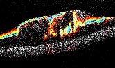
Imaging Modalities in the OCI Lab: Light and sound waves can be described by their degree of temporal coherence, i.e., how well one wave correlates with another at a later time. If the wave has low coherence, like the white light emitted from a light bulb, we can use it to perform optical depth ranging, a method that is called Optical Coherence Tomography (OCT). We use this method in our laboratory to build up 2- and 3-D images inside biological tissues. Analogously, we can use sound waves that have a broad bandwidth to perform ultrasonic imaging, which is based on the same depth-ranging principles.
We use both OCT and ultrasound in our laboratory. Ultrasound provides deeper penetration in tissues (several centimeters) but poorer resolution (hundreds of micrometers), which is complementary to OCT, which is more shallow (several millimeters) but higher resolution (several micrometers).
Tissue Viscoelastic Properties: Viscoelasticity is a mechanical property that describes both the elastic (rubber-like) and viscous (molasses-like) behavior of a material. Many human diseases are associated with changes in tissue viscoelasticity -- therefore, the ability to image these properties is very important in biomedical imaging. In our laboratory, we are investigating the viscoleastic properties in several systems, including the upper airway wall in its relation to sleep apnea and other obstructive disorders, airway mucus and its relevance to cystic fibrosis, early-stage breast cancer progression, and blood clots (thrombosis).
Developing New Methods for Imaging Contrast: In imaging science, contrast is the main way that we "see" the important patterns in an image. Often, biomedical images do not display the relevant properties of the tissue in order to diagnose or detect a disease. In our laboratory, we use several new methods for generating image contrast. This includes sensing the nanoporous structure of tissue from the diffusion rate of nanoparticle probes, detecting magnetic contrast agents using magnetomotive imaging, and measuring the intracellular motility of cells by their optical fluctuation patterns. Please see my research page for more details about our current research projects.
intro page - research - publications - people - open positions
UNC Physics & Astronomy - Biomedical Research Imaging Center - UNC Home